
Wounds
Like skin hemorrhaging, wounds represent breaks in the skin, a key barrier against external dangers such as pathogens. Unlike skin hemorrhaging, wounds are characterized by superficial or deeper damage to the epidermis, and in some cases, exposure of the subcutaneous tissue and muscles. Wounds can result from handling, contact with equipment, aggressive interactions, or disease. Beyond causing pain and stress, open wounds in direct contact with water can quickly become infected, posing a welfare risk.
| Level 0 | Level 1 | Level 2 | Level 3 |
|---|---|---|---|
|
No wounds |
Healed wounds |
Several small superficial wounds |
Many small wounds |
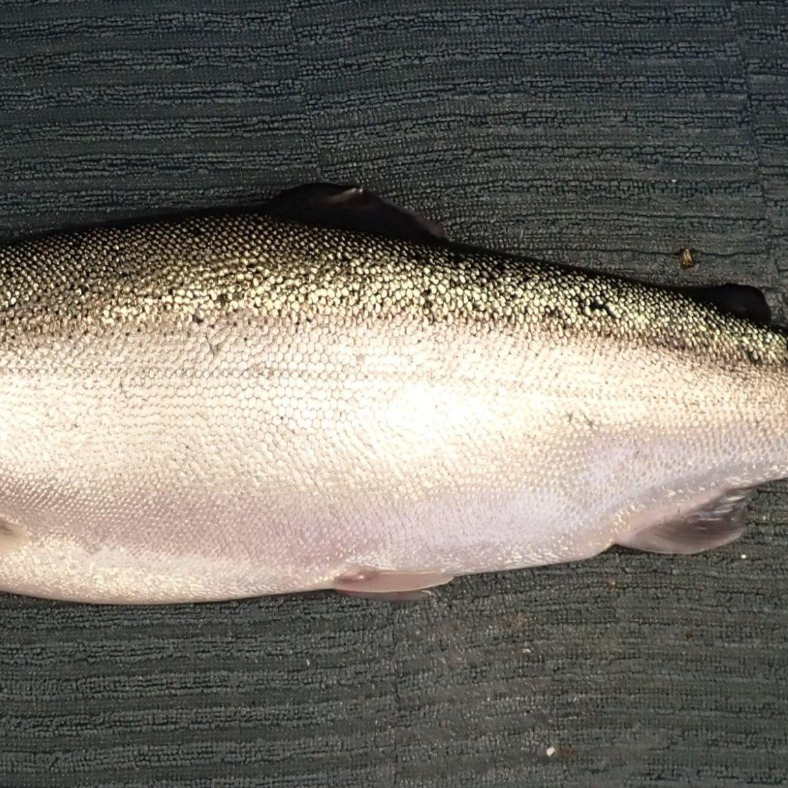
Healthy rainbow trout with no injuries97
|
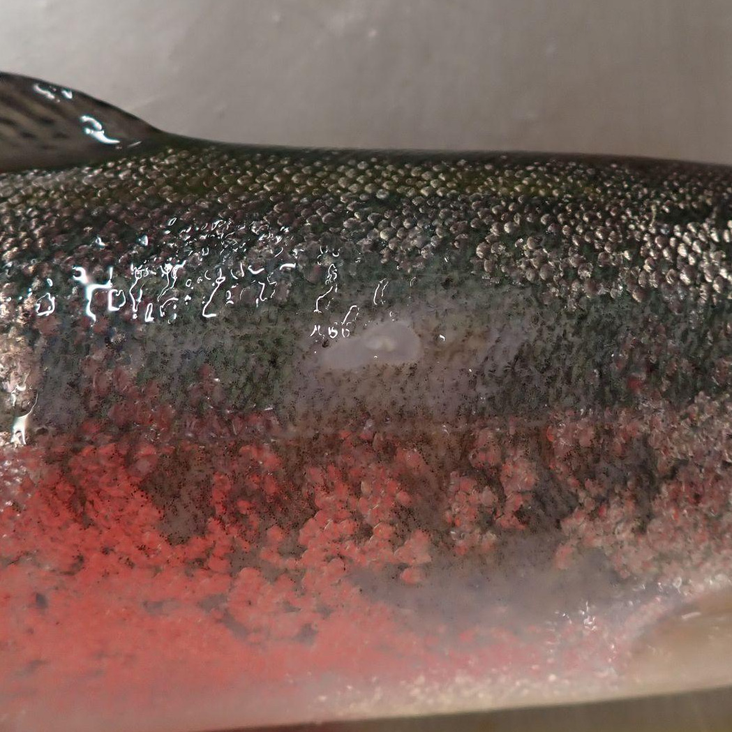
Rainbow trout with a small superficial wound98
|
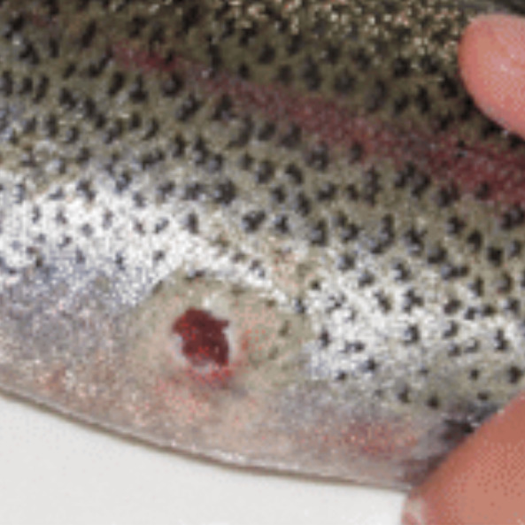
Rainbow trout with a wound99
|
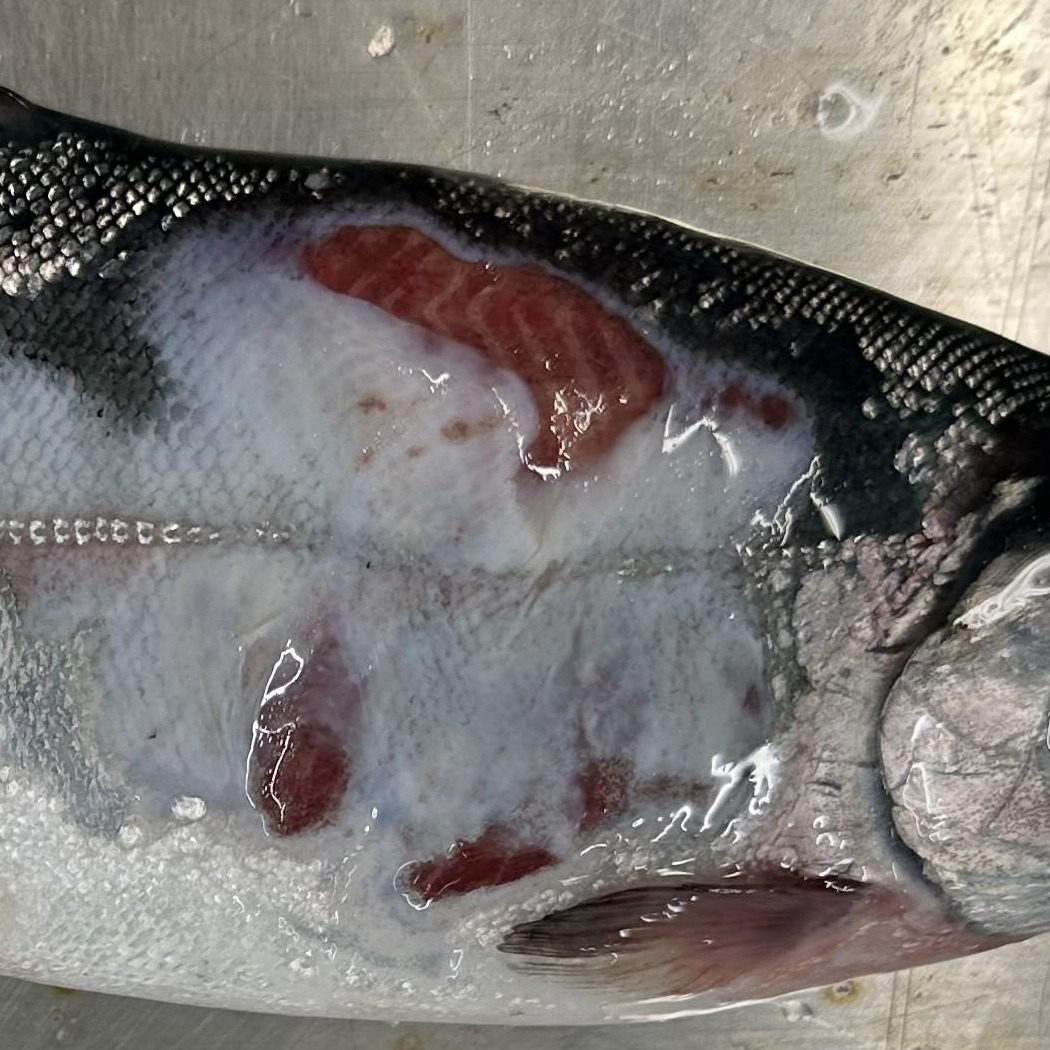
Rainbow trout with a large and deep wound100
|
Protocol application
Scoring this indicator involves counting total wounds, determining wound size (small or large), and checking the depth and infection status. This indicator addresses wounds on the body. To avoid double-counting, wounds on the fins or at the fin base are scored under fin damage, and wounds on the snout under snout damage.
Calculate the distribution of scores across the thresholds within the sample.
Scoring notes
- The status of injuries (fresh, healed, or infected) is critical to assess because fresh tissue damage is automatically more severe.
- Fresh wounds may have active bleeding, red or pink inflamed spots, potential swelling, or edema, and indicate recent trauma.
- Healed wounds have a stable size with scar tissue formed and no active bleeding, indicating the welfare issue has been largely resolved.
- Infected wounds include wounds with white or grey fungal growth, excessive debris or mucus, visible discharge, redness extending further than 0.5 cm from the wound edge, or necrotic tissue.
- Wound depth is also important to assess, as the deeper the wound is, the more severe its impact on welfare.
- A superficial wound is one where the muscle is not exposed. This includes wounds exposing the dermis or hypodermis.
- Wounds with any muscle exposed (or reaching the abdominal cavity) are automatically scored as level 3.
- Wound size classification is based on expert opinion, and inspired by guidance in FISHWELL.19 Therefore:
- Small wounds are visible only on close inspection.
- Moderate wounds are < 2 cm in diameter.
- Large wounds are ≥ 2 cm in diameter.
- For practical field reference, a UK one-penny coin, a 10-cent coin (Euro), or a US one-cent coin can be used to approximate this threshold.
- For smaller fish, it may be appropriate to use stricter thresholds to differentiate between a small wound and a large wound.
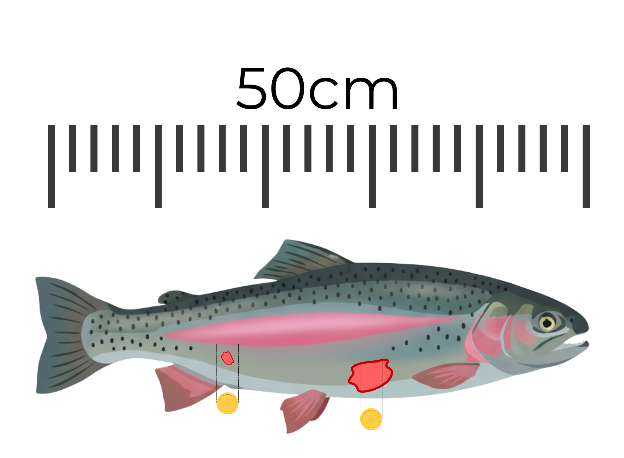
- Figure: Wound size reference on rainbow trout. The coin represents a 2cm diameter, approximating the threshold between a moderate wound (< 2cm) and a large wound (≥ 2cm).
- When scoring, note the wound location to further identify trends (e.g., dorsal surface, lateral sides, ventral surface, head).
- Validation: This scoring system has been adapted from the Laksvel scoring system for Atlantic salmon.
Key evidence
| Source | Summary of source | Key result |
|---|---|---|
| Schmidt et al., 2016 | Experimental study investigating wound healing in 154 rainbow trout (112 g) in freshwater laboratory conditions over 1 year. Four circular wounds were made between the lateral line and dorsal fin on the left side of each fish. The wounds were spaced 1cm apart, 3mm deep, and penetrated the muscle. Fish were divided between two tanks, with half in each tank wounded. In one of the tanks, the fish were exposed to an immunomodulatory β-glucan product. The temperature in the first 100 days was around 8.5°C. |
Inflammatory and matrix-remodeling genes were strongly upregulated from day 1 to at least day 14, and remained elevated until day 100. Wound areas enlarged until day 14 due to swelling and then slowly contracted. By day 100, the epidermis had closed, but the dermis remained thickened and disorganized, and only 20% of wounds had fully contracted. No proper muscle regeneration was identified. After one year, scales had not fully regenerated, with some small, irregular scales around wound edges. At this point, the wound appeared cosmetically healed. The immunomodulatory β-glucan treatment had no effect on wound closure speed. |
| Ingerslev et al., 2010 | Experimental study investigating the impacts of standardized skin and muscle injury on 80 rainbow trout (~43 g at time of injury, growing to ~84 g at the end of the experiment) in freshwater systems over 42 days. Each fish received approximately 50 punctures using a 25 needle array pressed twice in a 6mm x 6mm pattern to a 6 mm depth. The injury site was above the lateral line, on the left side, behind the dorsal fin. |
On days 7, 14, and 21, clear bleeding was visible, with swollen skin on day 7 and signs of inflammation still present but nearly resolved by day 14. By day 28, bleeding was no longer visible, and external swelling had essentially resolved. Expression of IL-1β and IL-8 (pro-inflammatory cytokines) increased post-injury but was localized to the site of injury. Upregulation decreased as the injury healed and was about normal at day 28. |
Supporting evidence
- Ingerslev, H. C., T. Lunder, and M. E. Nielsen. “Inflammatory and Regenerative Responses in Salmonids Following Mechanical Tissue Damage and Natural Infection.” Fish & Shellfish Immunology 29, no. 3 (September 2010): 440–50. https://doi.org/10.1016/j.fsi.2010.05.002.
- Nilsson, Jonatan, Kristine Gismervik, Kristoffer Vale Nielsen, Martin Haugsmo Iversen, Chris Noble, Jelena Kolarevic, Hilde Frotjold, et al. 2025. “Laksvel — a Standardised, Operational Welfare Monitoring Protocol for Atlantic Salmon Held in Sea Cages.” Institute of Marine Research. https://www.hi.no/en/hi/nettrapporter/rapport-fra-havforskningen-en-2025-40.
- Noble, Chris, Hernán A. Cañon Jones, Børge Damsgård, Matthew J. Flood, Kjell Ø. Midling, Ana Roque, Bjørn-Steinar Sæther, and Stephanie Yue Cottee. “Injuries and Deformities in Fish: Their Potential Impacts upon Aquacultural Production and Welfare.” Fish Physiology and Biochemistry 38, no. 1 (September 15, 2011): 61–83. https://doi.org/10.1007/s10695-011-9557-1.
- Noble, C., K. Gismervik, M. H. Iversen, J. Kolarevic, J. Nilsson, L. H. Stien, and J. F. Turnbull. 2020. “Welfare Indicators for Farmed Rainbow Trout: Tools for Assessing Fish Welfare.” Nofima. https://nofima.no/wp-content/uploads/2020/05/Welfare-Indicators-for-farmed-rainbow-trout-Noble-et-al.-2020.pdf.
- Pettersen, Jostein M., Marc B.M. Bracke, Paul J. Midtlyng, Ole Folkedal, Lars H. Stien, Håvard Steffenak, and Tore S. Kristiansen. “Salmon Welfare Index Model 2.0: An Extended Model for Overall Welfare Assessment of Caged Atlantic Salmon, Based on a Review of Selected Welfare Indicators and Intended for Fish Health Professionals.” Reviews in Aquaculture 6, no. 3 (June 30, 2013): 162–79. https://doi.org/10.1111/raq.12039.
- Schill, Daniel J., and F. Steven Elle. “Healing of Electroshock-Induced Hemorrhages in Hatchery Rainbow Trout.” North American Journal of Fisheries Management 20, no. 3 (August 1, 2000): 730–36. https://doi.org/10.1577/1548-8675(2000)020%3C0730:hoeihi%3E2.3.co;2.
- Schmidt, J.G., E.W. Andersen, B.K. Ersbøll, and M.E. Nielsen. “Muscle Wound Healing in Rainbow Trout (Oncorhynchus mykiss).” Fish & Shellfish Immunology 48 (January 2016): 273–84. https://doi.org/10.1016/j.fsi.2015.12.010.
- Stien, Lars H., Marc B. M. Bracke, Ole Folkedal, Jonatan Nilsson, Frode Oppedal, Thomas Torgersen, Silje Kittilsen, et al. “Salmon Welfare Index Model (SWIM 1.0): A Semantic Model for Overall Welfare Assessment of Caged Atlantic Salmon: Review of the Selected Welfare Indicators and Model Presentation.” Reviews in Aquaculture 5, no. 1 (March 2013): 33–57. https://doi.org/10.1111/j.1753-5131.2012.01083.x.
- Stien, Lars Helge, Chris Noble, Angelico Madaro, Jonatan Nilsson, René Alvestad, Kristine Gismervik, Ewa Harasimczuk, Hogne Bleie, Karoline Skaar Amthor, and Marc B. M. Bracke. “Reviewing the Severity of Handling-Induced Injuries in the LAKSVEL Protocol for Salmon Welfare.” Reviews in Aquaculture 17, no. 4 (August 14, 2025). https://doi.org/10.1111/raq.70076.
- Sveen, Lene Rydal “Aquaculture Relevant Stressors and Their Impacts on Skin and Wound Healing in Post-Smolt Atlantic Salmon (Salmo salar L.)” (PhD thesis, 2018), https://bora.uib.no/bora-xmlui/bitstream/handle/1956/19024/Dr.thesis_2018_Lene_Rydal_Sveen.pdf.
- Virtanen, Miiro Ilmari, Monica F. Brinchmann, Deepti Manjari Patel, and Martin Haugmo Iversen. “Chronic Stress Negatively Impacts Wound Healing, Welfare, and Stress Regulation in Internally Tagged Atlantic Salmon (Salmo salar).” Frontiers in Physiology 14 (April 3, 2023). https://doi.org/10.3389/fphys.2023.1147235.
-
Noble, Chris, Kristine Gismervik, Martin H. Iversen, Jelena Kolarevic, Jonatan Nilsson, Lars H. Stien, and James F. Turnbull. “Welfare Indicators for Farmed Atlantic Salmon: Tools for Assessing Fish Welfare.” Nofima, November 2018. https://nofima.no/wp-content/uploads/2021/05/FISHWELL-Welfare-indicators-for-farmed-Atlantic-salmon-November-2018.pdf.
-
Nilsson, Jonatan. 2026. Rainbow trout, score 0. Photograph. Personal communication, Institute of Marine Research.
-
Nilsson, Jonatan. 2026. Rainbow trout with a small superficial wound, score 1. Photograph. Personal communication, Institute of Marine Research.
-
Nilsson, Jonatan, Kristine Gismervik, Kristoffer Vale Nielsen, Martin Haugsmo Iversen, Chris Noble, Jelena Kolarevic, Hilde Frotjold, et al. 2025. “Laksvel — a Standardised, Operational Welfare Monitoring Protocol for Atlantic Salmon Held in Sea Cages.” Institute of Marine Research. https://www.hi.no/en/hi/nettrapporter/rapport-fra-havforskningen-en-2025-40.
-
Bui, Samantha. 2026. Rainbow trout with a large wound, score 3. Photograph. Personal communication, Institute of Marine Research.